Patent With Hepatopetal Flow In Fulton
Description


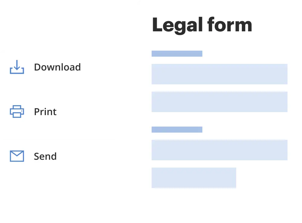
Get your form ready online
Our built-in tools help you complete, sign, share, and store your documents in one place.
Make edits, fill in missing information, and update formatting in US Legal Forms—just like you would in MS Word.
Download a copy, print it, send it by email, or mail it via USPS—whatever works best for your next step.
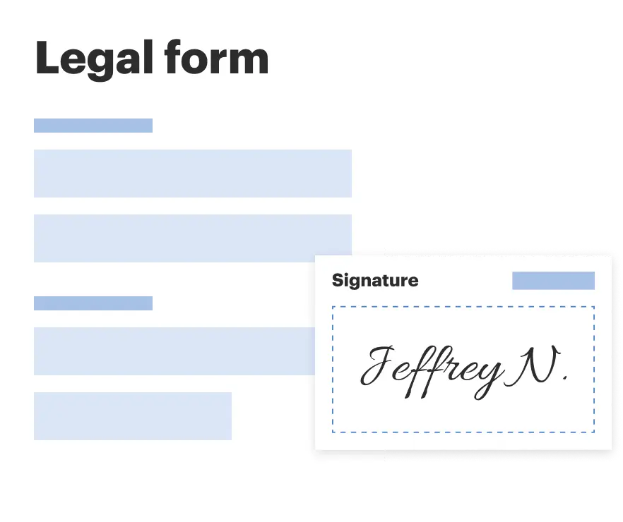
Sign and collect signatures with our SignNow integration. Send to multiple recipients, set reminders, and more. Go Premium to unlock E-Sign.
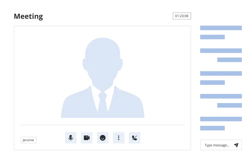
If this form requires notarization, complete it online through a secure video call—no need to meet a notary in person or wait for an appointment.
We protect your documents and personal data by following strict security and privacy standards.
Make edits, fill in missing information, and update formatting in US Legal Forms—just like you would in MS Word.
Download a copy, print it, send it by email, or mail it via USPS—whatever works best for your next step.
Sign and collect signatures with our SignNow integration. Send to multiple recipients, set reminders, and more. Go Premium to unlock E-Sign.
If this form requires notarization, complete it online through a secure video call—no need to meet a notary in person or wait for an appointment.
We protect your documents and personal data by following strict security and privacy standards.
Looking for another form?

Form popularity
FAQ
If the vein compresses, this confirms that the vein is open and patent. If the vein does not compress and debris is seen within the vein, this confirms the presence of a blood clot or obstruction of the vein accounting for the patient's symptoms.
Hepatopetal denotes flow of blood towards the liver, which is the normal direction of blood flow through the portal vein. The term is typically used when discussing the portal vein or recanalized vein of the ligamentum teres in patients with suspected portal hypertension. It is the opposite of hepatofugal.
The overall mortality rate of the patients with hepatofugal portal venous flow was 26.67% (95% confidence interval, 11.1%–42.9%). Causes of death were multiorgan failure with septic or hypovolemic shock (n = 5), hepatic failure (n = 2), and cardiac failure (n = 1).
A normal portal venous flow is hepatopetal. A flow reversal (or a hepatofugal flow) is seen in the case of portal hypertension (Fig. 6).
Hepatopetal denotes flow of blood towards the liver, which is the normal direction of blood flow through the portal vein. The term is typically used when discussing the portal vein or recanalized vein of the ligamentum teres in patients with suspected portal hypertension.
Hepatofugal or non-forward portal flow (NFPF) is an abnormal flow pattern in which the portal venous flow is from the periphery of the liver towards the porta hepatis and backwards along the portal vein. This phenomenon is not uncommon in patients with liver disease 3.
Patent track sign is a finding on color Doppler ultrasound, representing blood traveling along the course a biopsy needle track. It can occur after a biopsy of any organ, but is more often seen after liver or kidney biopsies.
Portal venous flow is approximately 1150 ml/min, as opposed to hepatic arterial flow, which is only 350 ml/min, resulting in total liver blood flow of 1500 ml/min. The portal vein supplies between 50 and 70% of the oxygen to the liver, especially during the fasting state.
Veins are very compressible or “collapsible”. Throughout the Ultrasound procedure, the sonographer will be gently pushing against the limb to compress the vein in that segment. If the vein compresses, this confirms that the vein is open and patent.