Motion Information Necessary With The Paralleling Technique
Description


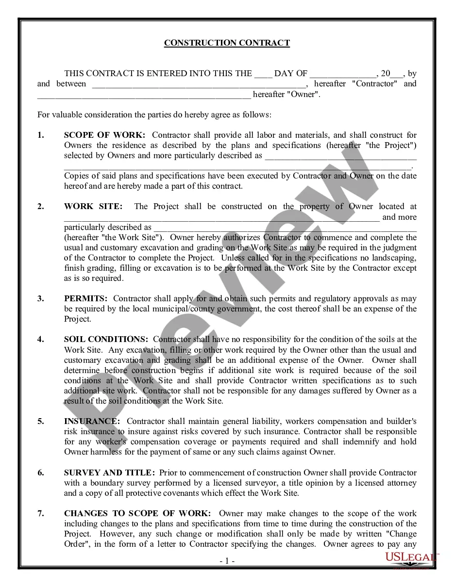
Get your form ready online
Our built-in tools help you complete, sign, share, and store your documents in one place.
Make edits, fill in missing information, and update formatting in US Legal Forms—just like you would in MS Word.
Download a copy, print it, send it by email, or mail it via USPS—whatever works best for your next step.
Sign and collect signatures with our SignNow integration. Send to multiple recipients, set reminders, and more. Go Premium to unlock E-Sign.
If this form requires notarization, complete it online through a secure video call—no need to meet a notary in person or wait for an appointment.
We protect your documents and personal data by following strict security and privacy standards.
Make edits, fill in missing information, and update formatting in US Legal Forms—just like you would in MS Word.
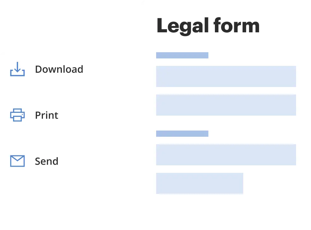
Download a copy, print it, send it by email, or mail it via USPS—whatever works best for your next step.
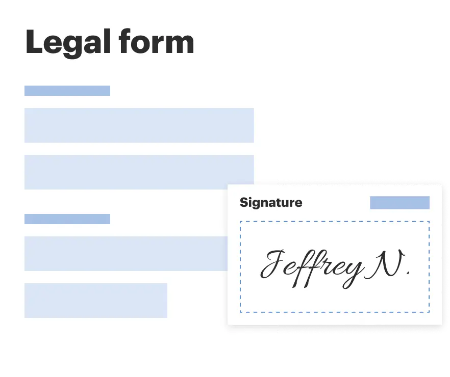
Sign and collect signatures with our SignNow integration. Send to multiple recipients, set reminders, and more. Go Premium to unlock E-Sign.
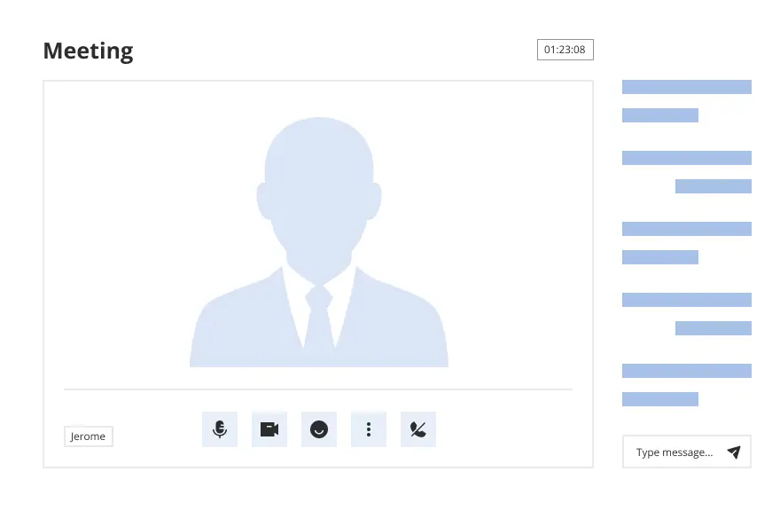
If this form requires notarization, complete it online through a secure video call—no need to meet a notary in person or wait for an appointment.

We protect your documents and personal data by following strict security and privacy standards.
Looking for another form?
How to fill out Motion For Discovery Of Information Necessary To Receive A Fair Trial?
Legal documents handling can be perplexing, even for seasoned experts. When you are seeking a Motion Information Required With The Paralleling Technique and lack the time to invest in searching for the correct and current version, the processes can be challenging.
An extensive online form repository could be a transformative solution for anyone who wishes to handle these matters effectively. US Legal Forms is a leading provider in online legal forms, with more than 85,000 state-specific legal documents accessible at any moment.
Save time and effort searching for the documents you require, and make use of US Legal Forms’ advanced search and Preview feature to find Motion Information Required With The Paralleling Technique and download it.
If you have a subscription, Log In to your US Legal Forms account, search for the form, and download it. Check your My documents tab to see the documents you have previously downloaded and to organize your folders as needed.
Take advantage of the US Legal Forms web library, supported by 25 years of expertise and reliability. Transform your everyday document management into a seamless and user-friendly experience today.
- Verify it is the correct form by previewing it and reviewing its description.
- Confirm that the sample is accepted in your state or county.
- Select Buy Now when you are ready.
- Choose a monthly subscription plan.
- Select the format you need, and Download, complete, sign, print, and send your document.
- Obtain access to state- or county-specific legal and business documents. US Legal Forms addresses any needs you may have, from personal to business paperwork, all in one location.
- Utilize cutting-edge tools to complete and manage your Motion Information Required With The Paralleling Technique.
- Access a valuable resource library of articles, guides, and manuals relevant to your case and requirements.
Form popularity
FAQ
The paralleling technique requires (1) that the film be placed parallel to the long axes of the teeth being radiographed and (2) that the x-ray beam be directed at right angles to both the film and long axes of the teeth.
With this technique, the film is placed parallel to the long axis of a tooth, allowing the X-ray to be focused perpendicular to the long axis of the tooth. The patient is seated upright in the dental chair and should remove any removable dental appliances, glasses or jewelry that could interfere with the X-ray beam.
Chat Image Receptor Placement. Position so that it will cover the correct teeth to be examined. Image Receptor Position. Positioned parallel to the long axis of the tooth. Vertical angulation. Central ray must be directed perpendicular to the image Receptor and the long axis of the tooth. Horizontal angulation. ... Central ray.
Answer: it helps position the film parallel to the long axis of the tooth and eliminates the need for the patient to stabilize the film.
Dental Assisting: Dental Radiology 1 - Parallel Technique - YouTube YouTube Start of suggested clip End of suggested clip Area instruct the patient to slowly bite down on the bite block. Slide the xep ring along the bar.MoreArea instruct the patient to slowly bite down on the bite block. Slide the xep ring along the bar. Until. It's as close to the patients face as possible position the tube head parallel.