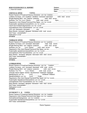
ROENTGENOLOGICAL REPORT Patient: ( ) Sent for Consult Doctor: Date: / / CERVICAL SPINE VIEWS .

How it works
  • Open form

    Open form follow the instructions

  • Easily sign form

    Easily sign the form with your finger

  • Share form

    Send filled & signed form or save

How to fill out the ROENTGENOLOGICAL REPORT Patient Sent For Consult online

This guide provides detailed instructions on completing the ROENTGENOLOGICAL REPORT Patient Sent For Consult form online. By following these steps, you will accurately fill out each section, ensuring a comprehensive report for the medical consult.

Follow the steps to successfully complete the form.

  1. Click ‘Get Form’ button to obtain the form and open it in the editor.
  2. Begin by filling in the patient's name in the 'Patient' field and the doctor's name in the 'Doctor' field. Make sure to include the date in the specified format.
  3. Move to the cervical spine section. Fill in the 'VIEWS' field with the appropriate imaging views taken. Indicate if there is a recent fracture in the visualized osseous structures and specify the location if applicable.
  4. Provide information about the lordotic curvature and select the applicable description (within normal limits, kyphotic, alordotic, increased, or decreased). Assess and mark the severity.
  5. For weight-bearing shift, select the appropriate option (within normal limits, anterior, or posterior) and mark the severity level.
  6. Indicate whether scoliosis is present, specifying if it is levo or dextro and noting the severity.
  7. Evaluate degenerative joint disease or narrowed disc space, providing levels if applicable. Repeat this process for hypertrophic spondylosis/osteoarthritis and facet/uncovertebral degeneration.
  8. Assess encroachment of the neuroforamina, indicating levels as needed. Move on to discuss the atlantodental interval (ADI) and provide the measurement in mm.
  9. Evaluate bone density and mark whether it is increased, adequate, or decreased, along with the severity level.
  10. In the soft tissue section, provide observations or mark it as unremarkable. Include any subluxation levels if applicable.
  11. Repeat steps 3 to 10 for the thoracic spine and lumbar spine sections, ensuring that all relevant information is accurately noted.
  12. For the extremity section, indicate views taken, recent fractures, degenerative joint disease, encroachment or impingement, and soft tissue observations.
  13. Finally, ensure that the doctor’s signature field is completed to validate the report.
  14. Once all sections are filled out correctly, save the changes, and then download, print, or share the completed form as necessary.

Complete your documents online today and ensure accurate submissions for better healthcare management.

Get form

Experience a faster way to fill out and sign forms on the web. Access the most extensive library of templates available.

Related content

Considerations for Exchanging and Sharing Medical...

Jun 28, 2016 — And rarely are the reports available with the images. ... When patients...

Learn more
5 Technology and Tools in the Diagnostic Process |...

Previous: 4 Diagnostic Team Members and Tasks: Improving Patient ... the report Health IT...

Learn more
LUX-Dx - Boston Scientific

Provide follow-up care for patients who have a LUX-Dx insertable cardiac monitor...

Learn more
Questions & Answers

Get answers to your most pressing questions about US Legal Forms API.

Contact support

What is a radiology report?

A detailed report that describes the results of an imaging test. A radiology report includes information about the type of imaging test that was done and how it was done. It also includes a brief medical history of the person having the test, including any symptoms or known diseases and why the test was needed.

Radiologist reporting performance cannot be perfect, and some errors are inevitable. Error or discrepancy in radiology reporting does not equate negligence. Radiologist errors occur for many reasons, both human- and system-derived.

A radiology report's "findings" section contains a detailed description and interpretation of the imaging findings observed during the examination. It provides a comprehensive assessment of the patient's anatomy, any abnormalities or pathologies detected, and their significance.

As a physician or an internist, we always look at the scans ourselves first before going through the radiologist's report or opinion. And this holds for any scans, be it CT, MRI or X-rays.

Indication. A radiology report's "indication" section provides a concise description of the clinical reason or suspicion that led to the ordering of the imaging study. It is a communication bridge between the referring healthcare provider and the radiologist interpreting the images.

Impression. In this section, the radiologist summarizes the findings and reports the most important findings that they see and possible causes (this is called a differential diagnosis) for those findings. This section offers the most important information for decision-making.

Using the diagnostic images, the radiologist or other providers can often: Diagnose the cause of your symptoms. Monitor how well your body is responding to a treatment you are receiving for your disease or condition. Screen for different illnesses, such as breast cancer, colon cancer, or heart disease.

Findings. This section lists what the radiologist saw in each area of the body in the exam. Your radiologist notes whether they think the area to be normal, abnormal, or potentially abnormal. Sometimes an exam covers an area of the body but does not discuss any findings.

Get This Form Now!

Use professional pre-built templates to fill in and sign documents online faster. Get access to thousands of forms.

If you believe that this page should be taken down, please follow our DMCA take down process here.

Get ROENTGENOLOGICAL REPORT Patient Sent For Consult