We use cookies to improve security, personalize the user experience, enhance our marketing activities (including cooperating with our marketing partners) and for other business use.
Click "here" to read our Cookie Policy. By clicking "Accept" you agree to the use of cookies. Read less
Read more
Accept
Loading
Form preview
  • US Legal Forms
  • Form Library
  • More Forms
  • More Uncategorized Forms
  • Microviewer Epithelial Lab 2006revised

Get Microviewer Epithelial Lab 2006revised

Name Date Period Epithelial Tissue Lab Microslides of animal tissue (set 50) Intro: There are 4 primary tissues: epithelial, connective, muscle and nervous. Epithelial tissue covers surfaces, lines.

How it works

  1. Open form

    Open form follow the instructions

  2. Easily sign form

    Easily sign the form with your finger

  3. Share form

    Send filled & signed form or save

How to fill out the Microviewer Epithelial Lab 2006revised online

Filling out the Microviewer Epithelial Lab 2006revised form can be an essential task for students studying epithelial tissue. This guide offers thorough instructions to help users navigate the form and complete it accurately, ensuring a clear understanding of the materials and concepts involved.

Follow the steps to efficiently complete the form online.

  1. Press the ‘Get Form’ button to access the Microviewer Epithelial Lab 2006revised form and open it in your browser.
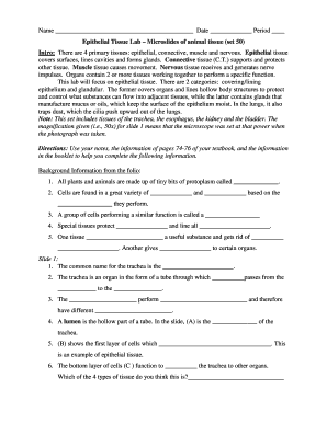
  2. Begin by entering your name on the designated line, followed by the date and the period of your class in the provided sections.
  3. Next, carefully review the introductory section about epithelial tissue and its functions, which will guide your responses in the following sections.
  4. Proceed to the background information section. Fill in the blanks by utilizing your class notes and relevant textbook pages. Ensure each answer aligns with the context of epithelial tissue.
  5. For Slide 1, answer questions regarding the common name for the trachea and its functions. Identify and label parts A, B, and C on the slide based on your knowledge of epithelial tissue.
  6. Move to Slide 2 and note the differences in magnification. Answer the questions and circle the correct representations of epithelial tissue.
  7. At Slide 3, engage in drawing and labeling the ciliated columnar epithelial cells as prompted. Use colors and labels to enhance clarity.
  8. Continue to Slide 4 to draw the simple cuboidal epithelium and respond to questions about gland arrangement and cell functionality.
  9. Skip ahead to Slide 6. Identify and describe the passage function. Complete the labeling and drawing task as instructed.
  10. Finish with Slide 7, drawing the simple squamous epithelium and comparing its thickness and characteristics to prior slides.
  11. Finally, ensure you save your changes, and download or print the completed Microviewer Epithelial Lab 2006revised form for submission.

Complete your Microviewer Epithelial Lab 2006revised form online today!

Get form

Experience a faster way to fill out and sign forms on the web. Access the most extensive library of templates available.
Get form

Related content

Adult Human Gingival Epithelial Cells as a Source...
Teeth develop from interactions between embryonic oral epithelium and ... teeth, providing...
Learn more
histology laboratory manual - Histology Lab Manual
This manual is a guide to work in the histology laboratory. ... Note the intense reaction...
Learn more
Adult Human Gingival Epithelial Cells as a Source...
Teeth develop from interactions between embryonic oral epithelium and ... teeth, providing...
Learn more

Related links form

FCC Screening Worksheet - Garretson Resolution Group CMS Proof Of Representation Form OR Executed Client Retainer ... Manpower Complement Sample HT Agt Form - Tsspdcl

Questions & Answers

Get answers to your most pressing questions about US Legal Forms API.

Contact support

If the epithelium is only one cell-layer thick, then it is called simple epithelium and if it is more than one layer thick, then it is called stratified epithelium. Finally, it is important to determine the shape of the apical cells (the cells at the free surface).

Functions as protection, diffusion, filtration, absorption, secretion, and sensory reception.

Epithelial tissues are widespread throughout the body. They form the covering of all body surfaces, line body cavities and hollow organs, and are the major tissue in glands. They perform a variety of functions that include protection, secretion, absorption, excretion, filtration, diffusion, and sensory reception.

Epithelial cells are a type of cell that lines the surfaces of your body. They are found on your skin, blood vessels, urinary tract, and organs. ... It's normal to have a small amount of epithelial cells in your urine.

Simple squamous epithelium This type of epithelia lines the inner surface of all blood vessels (endothelium), forms the wall of alveolar sacs in the lung and lines the body cavities (mesothelium). The primary function of simple squamous epithelia is to facilitate diffusion of gases and small molecules.

A squamous epithelial cell looks flat under a microscope. A cuboidal epithelial cell looks close to a square. A columnar epithelial cell looks like a column or a tall rectangle. A few epithelial layers are constructed from cells that are said to have a transitional shape.

Functions of epithelial cells include secretion, selective absorption, protection, transcellular transport, and sensing. Epithelial layers contain no blood vessels, so they must receive nourishment via diffusion of substances from the underlying connective tissue, through the basement membrane.

Epithelial tissues are identified by both the number of layers and the shape of the cells in the upper layers. There are eight basic types of epithelium: six of them are identified based on both the number of cells and their shape; two of them are named by the type of cell (squamous) found in them.

0:46 3:18 Suggested clip How to examine histology slides: tissues under a microscopeYouTubeStart of suggested clipEnd of suggested clip How to examine histology slides: tissues under a microscope

If there are squamous epithelial cells in your urine, it may mean your sample was contaminated. This means that the sample contains cells from the urethra (in men) or the vaginal opening (in women). It can happen if you do not clean well enough when using the clean catch method.

Get This Form Now!

Use professional pre-built templates to fill in and sign documents online faster. Get access to thousands of forms.
Get form
If you believe that this page should be taken down, please follow our DMCA take down processhere.

Industry-leading security and compliance

US Legal Forms protects your data by complying with industry-specific security standards.
  • In businnes since 1997
    25+ years providing professional legal documents.
  • Accredited business
    Guarantees that a business meets BBB accreditation standards in the US and Canada.
  • Secured by Braintree
    Validated Level 1 PCI DSS compliant payment gateway that accepts most major credit and debit card brands from across the globe.
Get Microviewer Epithelial Lab 2006revised
Get form
Form Packages
Adoption
Bankruptcy
Contractors
Divorce
Home Sales
Employment
Identity Theft
Incorporation
Landlord Tenant
Living Trust
Name Change
Personal Planning
Small Business
Wills & Estates
Packages A-Z
Form Categories
Affidavits
Bankruptcy
Bill of Sale
Corporate - LLC
Divorce
Employment
Identity Theft
Internet Technology
Landlord Tenant
Living Wills
Name Change
Power of Attorney
Real Estate
Small Estates
Wills
All Forms
Forms A-Z
Form Library
Customer Service
Terms of Service
Privacy Notice
Legal Hub
Content Takedown Policy
Bug Bounty Program
About Us
Blog
Affiliates
Contact Us
Delete My Account
Site Map
Industries
Forms in Spanish
Localized Forms
State-specific Forms
Forms Kit
Legal Guides
Real Estate Handbook
All Guides
Prepared for You
Notarize
Incorporation services
Our Customers
For Consumers
For Small Business
For Attorneys
Our Sites
US Legal Forms
USLegal
FormsPass
pdfFiller
signNow
airSlate WorkFlow
DocHub
Instapage
Social Media
Call us now toll free:
+1 833 426 79 33
As seen in:
  • USA Today logo picture
  • CBC News logo picture
  • LA Times logo picture
  • The Washington Post logo picture
  • AP logo picture
  • Forbes logo picture
© Copyright 1997-2025
airSlate Legal Forms, Inc.
3720 Flowood Dr, Flowood, Mississippi 39232