Loading
Get Microscope Diagram
How it works
-

Open form follow the instructions
-

Easily sign the form with your finger
-

Send filled & signed form or save
How to fill out the Microscope Diagram online
This guide provides step-by-step instructions to assist you in accurately completing the Microscope Diagram form online. By following these guidelines, you will ensure that all necessary components are properly documented.
Follow the steps to fill out the Microscope Diagram effectively.
- Click the ‘Get Form’ button to access the Microscope Diagram form and open it in your preferred online editor.
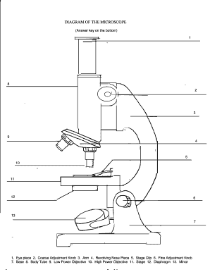
- Begin by locating the first component, the eye piece, and label it accordingly in the designated field.
- Proceed to the second component, the coarse adjustment knob, and enter its name in the corresponding field.
- Continue with the third component, the arm, following the same procedure for labeling.
- Move on to the revolving nose piece, entering its designation in the next section.
- For the fifth component, the stage clip, ensure it is accurately labeled.
- Identify and label the fine adjustment knob as the sixth component.
- Follow with the base as the seventh component, ensuring proper identification.
- Label the body tube as the ninth component in its appropriate field.
- Next, document the low power objective as the tenth component.
- Identify and label the high power objective as the eleventh component.
- Proceed to the stage and enter its designation as the twelfth component.
- For the thirteenth component, the diaphragm, ensure it is precisely labeled.
- Finally, enter ‘mirror’ in the designated field to complete the labeling of all components.
- Once you have completed labeling all sections, you can save changes, download, print, or share the form as needed.
Complete your Microscope Diagram online today!
To record from a microscope, first ensure that your setup is stable and properly focused. Use a camera or a smartphone to capture images or videos of your observations. Many microscopes offer a direct connection to computers, allowing you to save your studies digitally. This method creates a comprehensive microscope diagram that can serve as a valuable resource for future reference.